ap townes skull
AP axial skull Towne method 2. Long and narrow skull shape petrous ridges form an angle of 40 with the MSP internal structures positioned lower with reference to IOML -PA axial projection Caldwell -lateral -AP axial projection Townes What are the routine projections for.

Pin On Skulls
Angle CR 30 caudad if anterior clinoids are of primary interest.

. For patients unable to flex their neck align. Rest patients posterior skull against tableBucky surface. TOWNE METHOD skull series.
2-212 above the glabella. The CR exits the _____ _____ in the Townes skull. Depressed chin OML perpendicular to IR.
Patient is erect or supine. Occipital bone petrous pyramids and foramen magnum with dorsum sallae and posterior clinoids in its shadow are shown. Angle CR 37 caudad if dorsum sallae and posterior clinoid processes are of primary interest.
10 12 Grid Position Fig. Learn vocabulary terms and more with flashcards games and other study tools. Moving or stationary grid.
8-3 AP axial TowneCR 30 caudad to OML. The cross-table projection is a very effective alternative as the patient prefers to look up. 24 x 30 cm Portrait.
70 to 80 kV. The central ray is angled ____ degrees _____ in the Townes AP axial of the skull. Start studying AP axial Townes Skull.
AP PA Axial Skull AP Towne or PA Haas Method 24 30 cm LW. 100 - 115cm 40 inches. Ensure vertex of skull is within collimation field 4Ensure no rotation or tilt of headCENTERING.
Is the 10x12 cassette lengthwise or crosswise for an ap axial towne method Which position of the skull requires the IR to be crosswise lengthwise right or left lateral skull what position of the skull shows the petrous ridges over the lower 13 of the orbits pa 15 deg caldwell. Patient position and patient part. Regular CR and DR as recommended by manufacturer.
10 12 Grid Position Fig. Align MSP to CR and to midline of the table3. Skull foramen magnum.
Do image critique using PACEMAN guideline for the following projections. Align MSP to CR and to midline of the table3. Ensure vertex of skull is within collimation field 4Ensure no rotation or tilt of headCENTERING POINTANGULATIONOFTUBEAngle 30 degrees caudad to OML orAngle 37 degrees caudad to IOMLtrauma caseCenter at MSP 25 inches65cm above the glabella.
Soft Tissue Signs -Skull and Facial Bones. What is being demonstrated in the Townes skull. Skull - Townes Trauma Skull - Townes.
AP Axial Townes Skull CR Lines o _ Caudad OML _o Caudad IOML _ line perpendicular to IR _ Inches above glabella Right. No rotation or tilt Depress chin to bring OML or IOML perpendicular to IR. Center IR to projecting CR.
PA axial facial Caldwell method Expert Solution. No rotation or tilt Depress chin to bring OML or IOML perpendicular to IR. Flex neck to bring IOML perpendicular to midline of the grid or tableBucky surface.
AP PA Axial Skull AP Towne or PA Haas Method 24 30 cm LW. Seated erect or supine midsagittal plane aligned to CR and centerline perpendicular to IR. IR Size Orientation.
Seated erect or supine midsagittal plane aligned to CR and centerline perpendicular to IR. Skull - Hori Ray Lateral. IR size 10 x 12 24 x 30 cm.
8-3 AP axial TowneCR 30 caudad to OML. AP Skull no tube angle AP Skull no tube angle AP Axial Townes AP Axial Townes Projection Traditional lateral skull. This is an alternative projection for patients who cannot flex their neck sufficiently for AP axial Towne.
Center IR to projecting CR. View Skull positioningdocx from PA 0O at University of New England. Film Screen Combination.
PA axial facial Caldwell method Question. Where does the central ray enter in the Townes AP axial of the skull. Wednesday December 3 2014 HAAS METHOD SKULL SERIES Pathology Demonstrated.
AP axial skull Towne method 2. Fractures and pathologies of the skull. Head Clamps for Skull Radiography.
Skull - Townes Trauma - also called Skull - AP Axial Area Covered. PATIENT POSITIONRemove all metals plastics or other removable objects from the patients headPatient in erect or supinePART POSITION1. Up to 24 cash back The use of blocks and other radiolucent sponges will avoid exposing helpers hands to the primary beam and excluded as a film artifact.
Depress chin and make sure OML is perpendicular to IR 2. Depress chin and make sure OML is perpendicular to IR 2.

Skull Towne Method Ap Axial Projection Radiology Student Method Axial

Abdominal Radiographic Anatomy Wikiradiography Medical Radiography Radiology Student Medical Knowledge

Mountain Imaging Skull Positioning Radiology Schools Radiology Radiology Humor
Townes Skull Mp4 Radiology Technologist Book Worth Reading Medical

Picture Skull Mirror Selfie Caldwell

Pin On Radiographs

Pin On Radiographs

Standard Procedures For Skull Are Pa Caldwell Lateral Ap Axial Townes Radiology Imaging Radiology Student Radiography
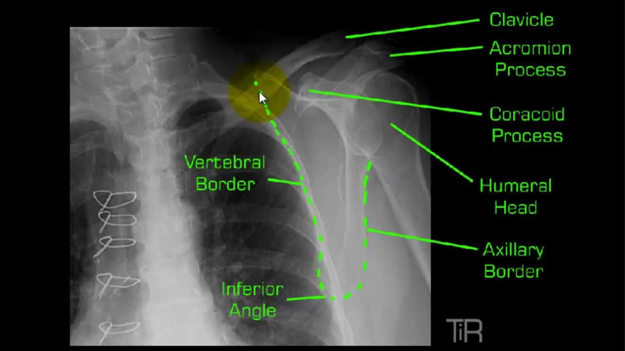
Y View Shoulder Mp4 Radiology Nursing Radiology Schools Anatomy

Pin On Radio Skull

Pin On Radiographs

Mandible Radiographic Anatomy Wikiradiography Facial Bones Radiology Medical Anatomy

Pin By Jamie L33 On Rad Tech Diagnostic Imaging Radiology Student Radiologic Technology

Pin On Radiology

Townes Skull Mp4 Radiology Technologist Book Worth Reading Medical

Cervical Spine Radiographic Anatomy Wikiradiography Diagnostic Imaging Radiology Radiology Schools

Wrist Radiographic Anatomy Wikiradiography Radiology Student Radiology Radiology Imaging

Punched Out Lesions In The Skull In A Case Of Multiple Myeloma Multiple Myeloma Myeloma Skull

Ap Skull Facial Bones Radiology Medical Radiography


Comments
Post a Comment